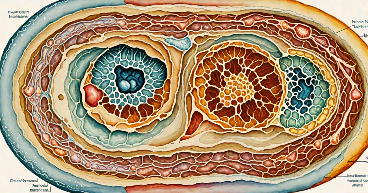
Human anatomy arises from a single fertilized egg through tightly regulated gene expression, cell differentiation, and morphogenesis during embryonic development. This process transforms undifferentiated cells into organized tissues, organs, and systems that function together.
At conception, a zygote forms from the fusion of sperm and egg. Within 24 hours, it divides into two cells, then four, eight, and so on. By day five, it becomes a blastocyst-a hollow ball of cells with an inner cell mass destined to form the body. Around day 14, gastrulation begins, forming three primary germ layers: ectoderm, mesoderm, and endoderm. These layers give rise to all major tissues and organs.
The ectoderm develops into the nervous system, skin, and hair. The mesoderm forms muscles, bones, kidneys, and the circulatory system. The endoderm gives rise to the lining of the digestive and respiratory tracts, liver, pancreas, and thyroid. This division is not arbitrary-it reflects evolutionary conservation across vertebrates. For example, the spinal cord and brain derive from the neural tube, which forms from the ectoderm's dorsal region.
Embryogenesis proceeds in phases. Weeks 3-8 are critical for organ formation. During week 4, limb buds appear. By week 6, the heart begins beating-its first contractions occur around day 21. Limb development follows a proximal-to-distal pattern: upper arm forms before forearm, then hand, then fingers. This sequence is controlled by signaling molecules like sonic hedgehog (SHH), which patterns digit identity.
By week 8, the embryo measures about 3 cm and resembles a miniature human. All major organs have initiated development. The face forms from six facial prominences that fuse at the midline. Failure in fusion leads to cleft lip or palate-conditions affecting approximately 1 in 700 births, according to the CDC.
After week 8, the term shifts from embryo to fetus. Growth continues, but now focus turns to size, maturation, and refinement. The fetal period sees rapid brain growth, myelination of nerves, and functional development of sensory systems. By birth, the average newborn weighs 3.4 kg and measures 50 cm-nearly 90% of adult height.
Human anatomy consists of interlocking systems, each derived from specific germ layers:
| System | Germ Layer | Key Organs | Developmental Timeline |
|---|---|---|---|
| Skeletal | Mesoderm | Bones, cartilage, joints | Weeks 6-12: ossification begins; long bones form via endochondral ossification |
| Nervous | Ectoderm | Brain, spinal cord, peripheral nerves | Week 4: neural tube closes; week 8: cerebral hemispheres begin folding |
| Circulatory | Mesoderm | Heart, blood vessels, blood cells | Week 5: heart tube loops; week 8: four-chambered structure established |
| Digestive | Endoderm | Liver, pancreas, intestines | Week 5: gut tube forms; week 10: liver produces blood cells |
Each system follows a precise timeline. For instance, the heart starts beating at 21 days post-fertilization, long before the brain shows electrical activity. This early cardiac function ensures nutrient delivery to the rapidly growing embryo.
Many assume anatomy is static-fixed at birth. But it changes continuously. Bone density peaks around age 30, then declines. The brain retains plasticity throughout life, adapting to injury or learning. Even the skeleton remodels itself every 10 years through osteoblast and osteoclast activity.
Another misconception is that anatomy results solely from genetics. While DNA provides the blueprint, environmental factors play a important role. Maternal nutrition affects fetal development-low folate intake increases neural tube defect risk by up to 70%, per the National Institutes of Health.
Some confuse anatomy with physiology. Anatomy describes structure-the "what." Physiology explains function-the "how." A muscle's anatomy includes fascicles, tendons, and motor neurons. Its physiology involves contraction mechanics, energy use, and fatigue response. Both are essential, but they're distinct fields.
Consider the leg. Mesoderm-derived somites segment along the body axis. Somitic cells migrate to the limb field and differentiate into skeletal muscle precursors. The limb bud grows under influence of SHH from the zone of polarizing activity (ZPA) at the posterior margin. This gradient determines digit identity-thumb forms with low SHH, pinky with high.
By week 8, the femur, tibia, fibula, and foot bones are visible. Cartilage models form first, then ossify. Blood vessels invade the cartilage matrix, bringing osteoblasts that deposit bone. The knee joint forms from synovial cavitation-fluid-filled spaces between opposing bones that later become joint capsules.
I once observed a 10-week human embryo specimen in a medical school lab. The limb buds were visible as small paddles. When viewed under a dissecting microscope, the vascular network was already forming in the webbing between digits. This confirmed that blood vessel development precedes digit separation-a detail often missed in simplified diagrams.
Understanding how human anatomy is made informs medicine, surgery, and regenerative science. Congenital disorders like spina bifida result from failed neural tube closure-prevented in many countries by folic acid supplementation before conception. Heart defects such as ventricular septal defects arise from improper septation during week 5-7.
Stem cell research aims to replicate developmental pathways. Scientists can now grow organoids-miniature, lab-grown versions of organs-from pluripotent stem cells. These models mimic early human development and are used to test drugs or study disease mechanisms without human trials.
For example, researchers at the University of Cambridge grew cerebral organoids that developed rudimentary eye-like structures. Though not functional vision, the presence of photoreceptor-like cells demonstrates how intrinsic developmental cues can drive complex organization even in vitro.
Learning how human anatomy is made reveals that the body isn't assembled piece by piece. It emerges through coordinated signals across time and space. Genes don't act alone-they interact with mechanical forces, chemical gradients, and cellular behaviors. A single mutation in the PAX6 gene can disrupt eye development entirely, leading to aniridia-a condition where the iris fails to form.
Understanding this process enables better diagnosis, treatment, and prevention of developmental disorders. It also guides advancements in bioengineering. The goal isn't to replace natural development-but to understand it well enough to support it when it goes awry.