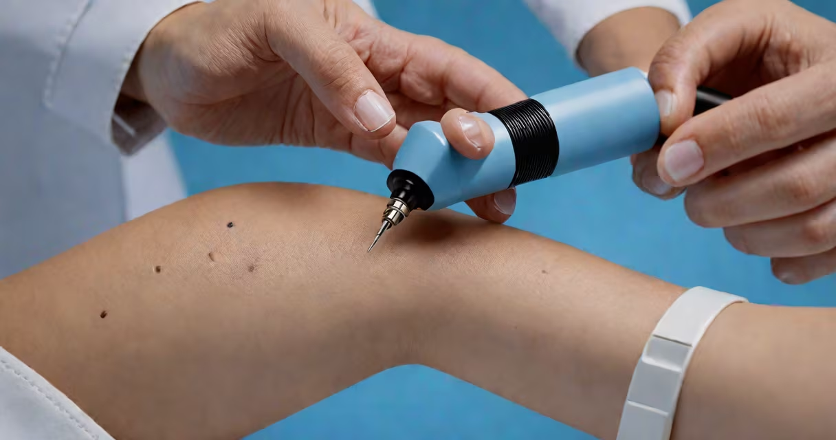
Stage 0 melanoma, also called melanoma in situ, is a non-invasive form of skin cancer where abnormal cells are present only in the epidermis-the outermost layer of the skin-and have not spread into deeper tissues or lymph nodes.
This stage represents the earliest detectable form of melanoma. It is not life-threatening when caught and treated promptly. The term "in situ" means the cancer remains localized and has not penetrated the basement membrane, which separates the epidermis from underlying layers.
According to the American Cancer Society, stage 0 accounts for about 10% of all newly diagnosed melanomas. It is typically identified during routine skin exams or when a suspicious mole is biopsied.
Stage 0 melanoma begins when pigment-producing cells (melanocytes) in the epidermis start dividing uncontrollably. These cells do not invade the dermis or travel through the bloodstream or lymphatic system.
The condition arises due to DNA damage in melanocytes, often caused by ultraviolet (UV) radiation from sunlight or tanning beds. Not all UV exposure leads to melanoma, but cumulative exposure increases risk. Genetic factors also play a role, especially in individuals with fair skin, red or blonde hair, and a history of sunburns.
A biopsy confirms stage 0 status. A pathologist examines tissue removed during a punch or excisional biopsy under a microscope. If cancer cells are found only in the epidermis and no invasion into the dermis is detected, the diagnosis is stage 0.
There are several subtypes of melanoma, and stage 0 can occur in any. The most common types seen at stage 0 include:
| Type | Description | Prevalence in Stage 0 |
|---|---|---|
| Superficial spreading melanoma | Spreads horizontally across the epidermis before invading downward. Most common subtype. | ~60% |
| Nodular melanoma | Aggressive growth pattern; often invades quickly. Rare in stage 0 but possible. | ~10% |
| Lentigo maligna melanoma | Develops in older adults on sun-exposed areas like the face. Often starts as a flat, pigmented lesion. | ~20% |
| Acral lentiginous melanoma | Rare; occurs on palms, soles, or under nails. Less likely to be stage 0 due to delayed detection. | ~5% |
Not all melanomas progress to invasive stages. Superficial spreading melanoma in stage 0 may remain stable for years if left untreated-but early removal prevents progression.
Stage 0 melanoma is usually discovered during a skin exam by a dermatologist. Suspicious moles are evaluated using the ABCDE rule:
If a lesion meets criteria, a biopsy is performed. A shave biopsy or punch biopsy removes a small sample of skin. Pathologists then analyze the tissue for signs of invasion.
One key diagnostic distinction: if the tumor shows no penetration below the basement membrane, it is classified as stage 0. If even minimal invasion into the dermis is detected, the stage advances to I.
Treatment for stage 0 melanoma is effective. The standard approach is surgical excision with narrow margins-typically 2-4 mm around the lesion.
For example, a patient presents with a 5 mm dark spot on their shoulder. Dermatologist performs a punch biopsy. Pathology report confirms melanoma in situ with no invasion. The lesion is surgically removed with 3 mm margins. Follow-up visits confirm complete healing with no recurrence.
Five-year survival rates for stage 0 melanoma exceed 99%. This high rate reflects the effectiveness of early intervention. Once melanoma invades deeper layers, survival drops.
According to the National Cancer Institute, patients diagnosed with stage 0 melanoma have a 98% five-year survival rate. Survival declines sharply after stage I, dropping to 92% at stage II and 70% at stage III.
Many people confuse stage 0 melanoma with benign moles or freckles. However, a mole that changes in appearance-even slightly-should be evaluated. Not all melanomas grow rapidly; some remain dormant for years.
A common error is delaying biopsy because the lesion appears small or harmless. But size does not correlate with severity. A lesion smaller than 1 mm can still be stage 0 melanoma.
Another misconception: stage 0 melanoma cannot spread. While it does not metastasize, it can progress to invasive disease if untreated. The American Academy of Dermatology warns that untreated stage 0 melanoma has a 10-15% chance of advancing to stage I within five years.
Any new or changing skin lesion should be assessed by a board-certified dermatologist. Early detection reduces the need for extensive treatment later.
I once observed a patient who ignored a small, dark spot on his forearm for two years. He finally sought care after it began bleeding. Biopsy confirmed stage 0 melanoma. After wide local excision, he required no further treatment. Had he waited another year, the cancer might have invaded the dermis.
After treatment, patients receive regular follow-ups. Surveillance includes annual skin checks and self-exams. The goal is to catch any recurrence early.
Stage 0 is not a permanent state. Without treatment, it can evolve into stage 1 melanoma-defined by invasion into the dermis, regardless of thickness.
Understanding the difference between stage 0 and stage 1 is critical. Stage 1 melanoma requires wider excision margins (usually 1-2 cm) and may involve sentinel lymph node biopsy in certain cases.
Stage 0 melanoma is treatable, curable, and preventable. Regular skin checks save lives. When caught early, it rarely progresses. The key is vigilance, prompt evaluation, and timely surgery.