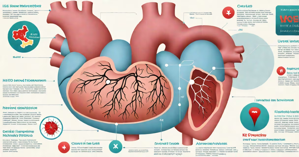
Ejection fraction (EF) is the percentage of blood pumped out of the left ventricle with each heartbeat. In heart failure, EF classifies the condition as either reduced (HFrEF) or preserved (HFpEF), guiding treatment decisions.
EF is measured using echocardiography, MRI, or nuclear scans. A normal EF ranges from 50% to 70%. When EF drops below 40%, it indicates heart failure with reduced ejection fraction (HFrEF), meaning the heart muscle is weak and cannot contract effectively. An EF of 50% or higher defines heart failure with preserved ejection fraction (HFpEF), where the heart contracts normally but stiffens and fails to fill properly.
Doctors use EF to distinguish between two major types of heart failure. HFrEF involves systolic dysfunction-weak pumping ability. HFpEF involves diastolic dysfunction-impaired filling despite normal contraction. The distinction affects medication choices, prognosis, and management strategies.
Standard echocardiograms calculate EF using the Simpson's method, which traces the left ventricle's volume during systole and diastole. The formula is: (end-diastolic volume - end-systolic volume) / end-diastolic volume × 100%. This yields a percentage representing the proportion of blood ejected per beat.
A patient with an end-diastolic volume of 150 mL and an end-systolic volume of 90 mL has an EF of (150 - 90) / 150 = 0.40, or 40%. This falls within the HFrEF range. A patient with similar volumes but lower end-systolic volume-say, 60 mL-would have an EF of 60%, indicating preserved function.
EF values are not static. They can fluctuate due to illness, medication changes, or recovery. A stable EF above 50% does not rule out heart failure. Some patients with HFpEF have EFs near 55% but still experience symptoms like shortness of breath and fatigue during exertion.
Heart failure is categorized by EF into two primary forms:
| Type | EF Range | Underlying Issue | Treatment Focus |
|---|---|---|---|
| Heart failure with reduced EF (HFrEF) | <40% | Systolic dysfunction-weak contraction | ACE inhibitors, beta-blockers, SGLT2 inhibitors, device therapy |
| Heart failure with preserved EF (HFpEF) | ≥50% | Diastolic dysfunction-stiff ventricle, poor filling | Blood pressure control, fluid management, treating comorbidities |
Despite similar symptoms, HFrEF and HFpEF differ in pathophysiology. HFrEF often results from prior myocardial infarction or cardiomyopathy. HFpEF is strongly linked to hypertension, diabetes, obesity, and aging. These differences explain why drugs effective in HFrEF-like carvedilol or sacubitril/valsartan-do not consistently improve outcomes in HFpEF.
One key misconception is that EF is the sole determinant of heart failure severity. A patient with an EF of 35% may feel better than another with 55% if the latter has advanced structural changes or comorbid conditions. EF is a tool, not a complete picture.
Many assume that a low EF always means severe disease. But EF can drop temporarily due to stress, infection, or arrhythmias. One patient I saw had an EF of 30% during a viral illness but recovered to 55% after treatment. His symptoms resolved, and he resumed normal activity. This shows EF must be interpreted alongside clinical context.
Another error is equating EF with symptom intensity. A person with EF of 45% may have debilitating fatigue and swelling, while someone with EF of 30% may be asymptomatic. Symptoms depend on neurohormonal activation, fluid overload, and organ perfusion-not just EF.
Some patients confuse EF with heart health. A high EF doesn't guarantee healthy coronary arteries or normal valve function. A patient with normal EF can still have significant aortic stenosis or ischemia. EF reflects pump efficiency, not vascular integrity.
EF guides diagnosis, risk stratification, and therapy selection. For HFrEF, evidence-based treatments reduce mortality by 30% or more. Drugs like sacubitril/valsartan and dapagliflozin are approved specifically for HFrEF based on trials like Model-HF and DAPA-HF.
In contrast, HFpEF lacks a proven mortality-reducing drug. Management focuses on controlling blood pressure, managing diabetes, reducing weight, and treating sleep apnea. The FDA has not approved any medication solely for HFpEF, though recent trials suggest potential benefit from finerenone in select populations.
EF also predicts hospitalization risk. Patients with EF below 35% face higher rates of rehospitalization and sudden cardiac death. This influences decisions about implantable cardioverter-defibrillators (ICDs) and cardiac resynchronization therapy (CRT).
During a clinic visit, I reviewed a 68-year-old woman with dyspnea on exertion. Her EF was 52%, but her echocardiogram showed thickened walls and elevated filling pressures. She met criteria for HFpEF. Despite normal EF, she responded well to spironolactone and lifestyle changes. Her case illustrates that EF alone cannot define heart failure.
EF is not the only measure of cardiac performance. Global longitudinal strain (GLS)-assessed via speckle-tracking echocardiography-detects subtle wall motion abnormalities before EF declines. GLS can identify early dysfunction in athletes or patients with chemotherapy exposure.
EF also differs from stroke volume (SV), which measures absolute blood volume ejected per beat. SV is influenced by heart rate and preload. Two patients with identical EFs can have vastly different SVs depending on size and heart rate. A 200-pound man with a resting heart rate of 60 beats per minute may have a higher SV than a 120-pound woman with the same EF.
EF is not interchangeable with cardiac output-the total volume of blood pumped per minute. Cardiac output = heart rate × stroke volume. A patient with low EF may maintain adequate cardiac output through tachycardia, masking underlying impairment.
Understanding EF requires knowing its limits. It is a snapshot of systolic function, not a full assessment of heart health. Always interpret EF in context with symptoms, imaging findings, and comorbidities.
Knowing what EF means in heart failure clarifies diagnosis and treatment. It is not a standalone verdict but a critical piece of a larger clinical puzzle.