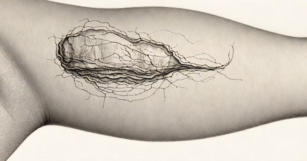
A bulla on the skin is a fluid-filled blister measuring 5 millimeters or more in diameter. It appears as a raised, sac-like lesion containing clear, serous, or bloody fluid. Bullae form when fluid accumulates between layers of the epidermis due to inflammation, trauma, or disease.
Bullae differ from smaller blisters called vesicles, which are less than 5 mm wide. The size threshold is clinically significant because it influences diagnosis and treatment. A bulla may develop rapidly or persist for days, depending on the underlying cause. They often appear on areas exposed to friction, burns, or allergic reactions, but can also signal systemic illness.
According to the American Academy of Dermatology Association, bullae are common in conditions such as pemphigus vulgaris, bullous pemphigoid, and toxic epidermal necrolysis. They are not always painful-some are asymptomatic-but they carry a risk of infection if ruptured. Early recognition improves outcomes, especially in autoimmune and infectious skin disorders.
Bullae arise when fluid builds up beneath the epidermis, typically between the stratum spinosum and stratum granulosum. This space is normally sealed by tight junctions. When those barriers break-due to immune attack, chemical exposure, or physical stress-fluid leaks into the space and forms a blister.
The fluid inside a bulla is usually serous (clear plasma-like fluid), but it can be cloudy, bloody, or purulent depending on the cause. Bloody bullae suggest trauma or vasculitis. Purulent fluid indicates infection. Serous bullae are common in autoimmune diseases like bullous pemphigoid.
For example, in a patient with suspected pemphigus vulgaris, a biopsy of a bulla shows acantholysis-the separation of keratinocytes due to autoantibodies attacking desmoglein proteins. This finding confirms the diagnosis and guides treatment with immunosuppressants.
Bullae fall into categories based on cause: traumatic, inflammatory, infectious, and autoimmune. Each type presents differently and requires distinct management.
| Type | Common Causes | Appearance | Typical Location |
|---|---|---|---|
| Traumatic | Burns, friction, pressure | Clear fluid, may be tender | Feet, hands, pressure points |
| Infectious | Herpes simplex virus, impetigo | Cloudy or yellow fluid, may crust | Face, mouth, genital area |
| Autoimmune | Bullous pemphigoid, pemphigus vulgaris | Large, flaccid, may rupture easily | Trunk, flexural areas |
| Inflammatory | Stevens-Johnson syndrome, toxic epidermal necrolysis | Widespread, painful, may peel | Body-wide, often triggered by drugs |
One non-obvious distinction: bullae in herpes infections tend to cluster and rupture quickly, forming shallow erosions. In contrast, bullae from bullous pemphigoid are tense, slow to rupture, and often appear in skin folds like the groin or axilla. Misdiagnosis can delay treatment-especially in elderly patients who may not report pain but show widespread bullae.
I once evaluated a 72-year-old man with large, painless bullae on his arms and legs. Initial suspicion was contact dermatitis. But when the lesions didn't improve with steroids, a skin biopsy revealed linear IgG deposits along the basement membrane-confirming bullous pemphigoid. He responded well to dapsone and prednisone within two weeks.
Bullae are frequently mistaken for other skin lesions. A common error is confusing bullae with nodules or cysts. Nodules are solid, deep-seated lumps; bullae are fluid-filled and superficial. Another confusion arises between bullae and large vesicles-both are fluid-filled, but size defines the difference. Vesicles are under 5 mm; bullae are 5 mm or greater.
Some assume all bullae require drainage. That's incorrect. As the American Academy of Dermatology notes, draining a bulla increases infection risk and delays healing. Intact bullae act as natural dressings. Only drain if the blister is very large, painful, or likely to burst during movement.
Another misconception: bullae always indicate serious illness. While some are signs of life-threatening conditions like toxic epidermal necrolysis, many result from minor causes-like a new pair of shoes causing friction blisters. Still, persistent or recurring bullae warrant evaluation, especially if accompanied by fever, joint pain, or mucosal involvement.
See a dermatologist if a bulla lasts longer than seven days, spreads rapidly, or appears with systemic symptoms like fatigue, weight loss, or fever. Bullae on mucous membranes (mouth, eyes, genitals) are red flags for autoimmune disease or drug reactions.
Diagnosis begins with visual inspection and history. Doctors assess location, number, fluid color, and associated symptoms. If the cause is unclear, they perform a skin biopsy with direct immunofluorescence. This test detects antibodies bound to skin layers-critical for diagnosing pemphigus or bullous pemphigoid.
For example, a patient with generalized bullae and a history of amoxicillin use had a biopsy showing IgG deposition in the dermal-epidermal junction. The diagnosis was drug-induced bullous eruption. Discontinuing the medication led to resolution within three weeks.
Testing for infections like herpes simplex virus involves PCR swab testing from the blister base. Blood tests may check for autoimmune markers such as anti-desmoglein antibodies. Imaging is rarely needed unless internal organs are involved.
A bulla on the skin is a fluid-filled blister 5 mm or larger. It signals a range of conditions-from minor trauma to severe autoimmune disease. Size matters: bullae are distinct from smaller vesicles. Not all require treatment, but persistent or widespread bullae demand medical attention. Early diagnosis prevents complications like scarring, infection, or progression to systemic illness.