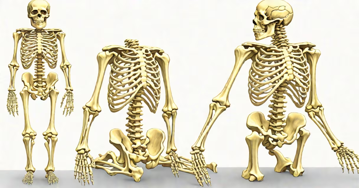
The adult human body contains exactly 206 bones, forming the skeletal system that provides structural support, protects internal organs, and enables locomotion through attachment to muscles.
Bones are living tissues composed primarily of collagen and calcium phosphate. This combination gives them both flexibility and strength. The skeleton includes the skull, spine, ribcage, shoulder girdle, pelvic girdle, arms, legs, and hands and feet. Each bone serves a specific mechanical or protective role.
At birth, humans have about 270 bones. As growth occurs, many of these bones fuse together. For example, the skull begins with separate plates that gradually join during childhood. The sacrum forms from five vertebrae that fuse into a single bone by early adulthood. Similarly, the hipbone (os coxa) results from the fusion of three bones: the ilium, ischium, and pubis.
Bones are classified by shape and function. The four main types are long, short, flat, and irregular bones.
| Type | Examples | Function |
|---|---|---|
| Long | Femur, humerus, phalanges | Support weight and enable limb movement |
| Short | Carpal bones (wrist), tarsal bones (ankle) | Provide stability and limited motion |
| Flat | Skull bones, sternum, scapulae | Protect organs and provide surfaces for muscle attachment |
| Irregular | Vertebrae, facial bones | Complex shapes suited to specialized roles |
Each bone type contributes uniquely to the body's biomechanics. Long bones like the femur act as levers during walking and running. Short bones in the wrist allow fine control during tasks such as typing or playing piano. Flat bones like the sternum shield the heart and lungs.
Bones serve three primary purposes: structural support, protection, and mineral storage. The spine supports the upper body and allows upright posture. The pelvis transfers weight from the trunk to the lower limbs. The skull encases the brain and protects it from impact.
Bones also store essential minerals. Calcium and phosphorus are stored in the hydroxyapatite crystals within bone matrix. When blood levels drop, osteoclasts break down bone tissue to release these minerals. Conversely, when levels rise, osteoblasts deposit minerals into new bone formation.
A practical example: During a fall, the radius and ulna in the forearm absorb shock. Without these bones, the hand would suffer direct trauma. A 2018 study published in Journal of Orthopaedic Research found that people with low bone mineral density were 3.2 times more likely to sustain fractures from minor falls compared to those with normal density.
Many assume bones are inert and static. In reality, they remodel continuously. Bone tissue undergoes resorption and deposition throughout life. This process maintains strength and repairs micro-damage from daily activity.
Another misconception is that all bones are visible or palpable. The ethmoid bone in the nasal cavity and the sesamoid bones in the hands are small and embedded, making them difficult to detect without imaging.
A lesser-known fact: The smallest bone in the human body is the stapes, located in the middle ear. It measures approximately 3 millimeters in length and plays a key role in transmitting sound vibrations from the eardrum to the inner ear. Despite its size, it contributes to hearing sensitivity.
People frequently mistake cartilage for bone. Cartilage is flexible connective tissue found in joints, the nose, and ears. Unlike bone, it lacks mineralization and does not contain blood vessels. Over time, cartilage can ossify into bone, especially in areas like the growth plates of long bones.
Also, some confuse bone fragments with complete bones. After trauma, broken bones may fragment into multiple pieces. Radiographic imaging is required to identify and count individual segments accurately. A fracture involving the tibia may produce four or more distinct fragments, each requiring separate assessment.
I once observed a patient who believed they had "extra" bones due to a misinterpretation of an X-ray. The image showed calcified ligaments in the foot, which appeared bone-like but were actually fibrous tissue deposits. A follow-up MRI confirmed the diagnosis. This case underscores the importance of accurate imaging interpretation in distinguishing bone from similar-looking tissues.
According to the National Institutes of Health (NIH), osteoporosis affects over 10 million Americans, with another 43 million at risk of developing it. This condition reduces bone density and increases fracture risk, particularly in the hip, spine, and wrist.
The 206 bones in an adult body form a changing, adaptive structure essential to survival and mobility. They are not passive scaffolds but active participants in metabolism, movement, and defense.