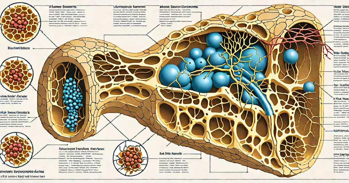
Lymph nodes are small, bean-shaped organs distributed throughout the lymphatic system that filter lymph fluid and host immune cells responsible for detecting and responding to pathogens, cancer cells, and foreign substances. They contain specialized compartments housing B cells, T cells, dendritic cells, macrophages, and stromal cells organized into distinct zones.
Each lymph node functions as a surveillance hub. When infection or abnormal cell activity occurs, antigen-presenting cells carry debris from tissues to the node via afferent lymphatic vessels. Once inside, these antigens are processed and presented to lymphocytes, initiating adaptive immune responses. The entire process begins within minutes of pathogen entry and can generate a measurable immune reaction in under 24 hours.
According to the National Institutes of Health (NIH), lymph nodes contain approximately 10^6 to 10^7 lymphocytes per gram of tissue, making them among the most densely populated immune sites in the body. This high concentration enables rapid detection and response to threats.
Lymph nodes receive unfiltered lymph through multiple afferent vessels entering the convex side. The fluid flows through sinusoids-narrow channels lined with macrophages-before reaching the medulla. As it moves, pathogens and particulate matter are trapped and destroyed by phagocytic cells.
Antigen-specific activation occurs primarily in the cortex. B cells cluster in follicles, while T cells reside in paracortical zones adjacent to high endothelial venules (HEVs). These HEVs allow circulating naive T cells to enter the node and scan for antigen-presenting dendritic cells. When a match occurs, T cells proliferate and differentiate into effector cells.
A key event is the formation of germinal centers-transient structures that develop within follicles during active immune responses. Here, B cells undergo somatic hypermutation and class switching, producing high-affinity antibodies tailored to specific antigens. This process takes about 5-7 days and results in long-lived plasma cells and memory B cells.
The internal architecture of a lymph node is divided into functional regions, each with a unique cellular composition:
| Region | Primary Cells | Function |
|---|---|---|
| Follicles (primary/secondary) | B cells, follicular dendritic cells | Initiate humoral immunity; form germinal centers during antigen exposure |
| Paracortex | T cells, dendritic cells | Site of T-cell activation and proliferation; supports cell-to-cell signaling |
| Medullary cords and sinuses | Macrophages, plasma cells, some B cells | Filter lymph; eliminate debris; secrete antibodies into efferent lymph |
| Subcapsular sinus | Macrophages, dendritic cells | First site of antigen capture from incoming lymph |
Stromal cells-fibroblastic reticular cells (FRCs)-form a scaffold that guides immune cell migration. FRCs produce chemokines like CCL19 and CCL21, which direct T cells and dendritic cells toward T-cell zones. Disruption of this network impairs immune coordination, as shown in mouse models where FRC depletion led to delayed pathogen clearance.
One non-obvious fact: lymph nodes do not produce lymphocytes. All B and T cells originate in bone marrow and thymus, respectively, then migrate to lymph nodes to mature and respond. The node itself acts as a training ground, not a factory.
Many assume lymph nodes swell because they are "fighting" infection directly. In reality, swelling results from increased cell proliferation and fluid accumulation-not from active combat. The enlargement reflects immune system activation, not defensive effort.
Another misconception is that all lymph nodes serve the same purpose. In fact, their location determines function. Cervical nodes drain the head and neck; axillary nodes handle upper limb and breast tissue; inguinal nodes monitor lower limbs and pelvic regions. A swollen inguinal node may indicate a sexually transmitted infection, while a cervical node enlargement often signals a sore throat or dental abscess.
For example, a patient with streptococcal pharyngitis typically develops tender, enlarged cervical lymph nodes within 24-48 hours. Histological analysis shows expanded germinal centers filled with activated B cells and plasma cells. This confirms that the node is processing antigen from the infected tonsil, not merely reacting passively.
Not all lymph node swelling indicates serious illness. However, persistent enlargement (>2 weeks), painless growth, or hard, fixed nodes warrant evaluation. The American Cancer Society notes that lymphadenopathy lasting longer than four weeks has a 10-15% chance of being due to malignancy, especially in adults over 40.
Conditions such as tuberculosis, HIV, sarcoidosis, and lymphoma can mimic reactive hyperplasia. Differentiation requires imaging (ultrasound or CT), fine-needle aspiration biopsy, or excisional biopsy. A single node larger than 1 cm in diameter in a non-infected area raises concern.
I once observed a case where a 52-year-old man presented with a 2.5 cm left-sided cervical node. Initial tests showed no signs of infection. A biopsy revealed diffuse large B-cell lymphoma. The node's size and lack of tenderness were red flags missed in early assessment. This underscores that internal structure-not just appearance-determines clinical significance.