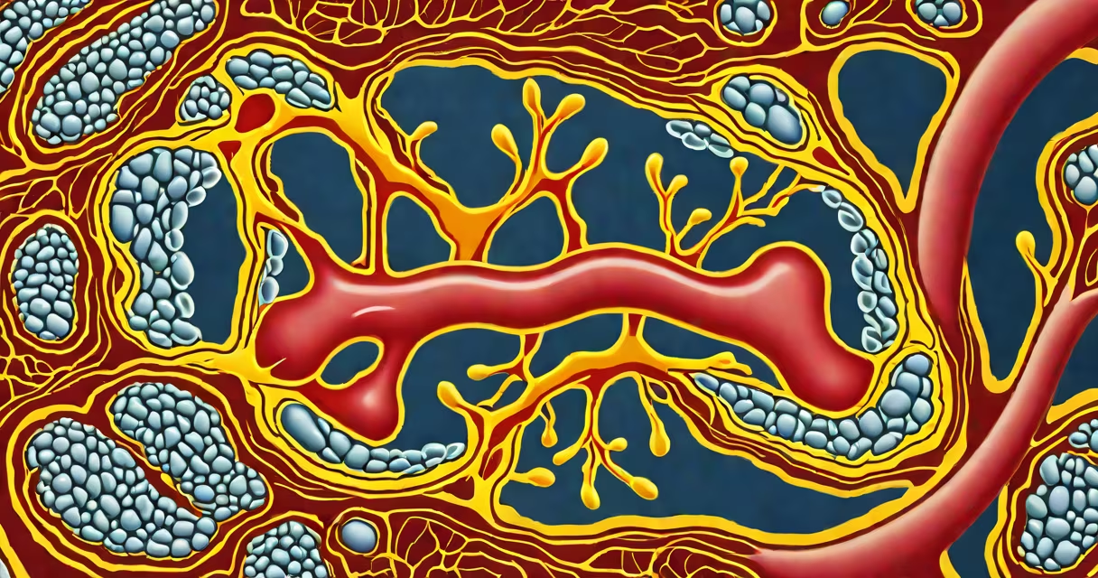
Atherosclerosis calcification of the aorta is the buildup of calcium within the walls of the aorta due to advanced atherosclerotic plaque. This process stiffens the vessel, reduces elasticity, and increases cardiovascular risk. It's detectable on imaging and associated with higher rates of heart attack and stroke.
Calcification occurs when calcium salts deposit in damaged arterial tissue. In the aorta-the largest artery carrying blood from the heart-this happens over years as cholesterol-rich plaques accumulate and trigger inflammation. Immune cells enter the vessel wall, release signaling molecules, and attract calcium crystals. Once deposited, calcium cannot be removed naturally. The result is a rigid, thickened aortic segment that fails to expand and contract properly during each heartbeat.
According to the American Heart Association, calcified aortic plaques are a key marker of systemic atherosclerosis. They indicate widespread vascular damage, not just localized disease. The presence of calcium in the aorta correlates strongly with mortality from cardiovascular events. Even in asymptomatic individuals, detection via coronary CT angiography shows increased risk of future cardiac events.
Calcification begins with endothelial injury-damage to the inner lining of the aorta. Causes include high blood pressure, elevated LDL cholesterol, smoking, and diabetes. When the endothelium is compromised, low-density lipoprotein (LDL) particles seep into the vessel wall and oxidize. Oxidized LDL triggers monocytes to migrate into the intima, where they become macrophages and engulf the lipid material. These cells turn into foam cells, forming fatty streaks-the earliest sign of atherosclerosis.
Over time, smooth muscle cells proliferate, and the lesion grows into a fibrofatty plaque. As the plaque matures, it undergoes necrosis and releases cellular debris. Calcium ions bind to phospholipids and proteins in the extracellular matrix, forming hydroxyapatite crystals. These crystals grow and aggregate, leading to visible calcification on imaging. The process resembles bone formation, though it occurs in soft tissue. This is known as metastatic calcification, driven by inflammatory signals rather than normal bone metabolism.
Once calcium deposition reaches a threshold, it becomes detectable on non-contrast CT scans. The Agatston score-a standard measure-quantifies calcium burden based on density and area. A score above 100 indicates moderate to severe calcification. Research published in Circulation shows patients with aortic calcium scores over 400 have a 3.5-fold higher risk of major adverse cardiac events over five years compared to those below 100.
Calcification in the aorta typically appears in three patterns:
| Type | Description | Common Location | Associated Risk |
|---|---|---|---|
| Intimal | Calcium deposits within the innermost layer of the aorta, aligned with plaque growth | Thoracic and abdominal aorta | High-linked to plaque instability and rupture risk |
| Medial | Calcium in the middle layer (media), often seen in aging or metabolic disorders | Abdominal aorta, especially near renal arteries | Moderate-associated with reduced compliance and hypertension |
| Perivascular | Calcium around the aorta, not inside the wall | Surrounding aortic branches | Low-often incidental, may reflect systemic calcium imbalance |
Imaging modalities differ in sensitivity. Non-contrast CT detects calcium with 95% accuracy. Ultrasound can identify calcification in the abdominal aorta but misses early deposits. MRI lacks specificity for calcium. CT remains the gold standard for quantifying calcification burden.
During a routine chest CT scan for lung nodule evaluation, a radiologist noticed dense calcifications along the posterior wall of the descending thoracic aorta in a 67-year-old man with hypertension and hyperlipidemia. The Agatston score was 620. He had no symptoms. Over the next three years, he developed angina and underwent coronary angiography, revealing 70% stenosis in the left anterior descending artery. His aortic calcification predicted the extent of coronary disease.
Many assume calcification is simply "hardening" caused by poor diet. While nutrition plays a role, calcification is a complex biological process involving immune response, mineral regulation, and vascular remodeling. It is not reversible through diet alone. Removing calcium deposits surgically is not feasible due to location and risk of rupture.
Another myth is that calcification only affects older adults. While prevalence rises with age-over 50% of people over 70 show some aortic calcification-it can appear earlier in individuals with chronic kidney disease or genetic disorders like pseudogout. A study in The Journal of the American College of Cardiology found 12% of patients under 50 with familial hypercholesterolemia had detectable aortic calcification.
Some confuse aortic calcification with aortic aneurysm. An aneurysm involves dilation and thinning of the vessel wall, while calcification involves thickening and stiffening. Both increase rupture risk but through different mechanisms. Aneurysms are monitored for size; calcification is monitored for progression and association with other cardiovascular events.
Calcification of the aorta is not a standalone diagnosis. It's a biomarker of advanced vascular disease. Its presence signals that plaque has progressed beyond early stages and that systemic inflammation is active. This makes it a useful predictor of future cardiac events, even when cholesterol levels are controlled.
I've reviewed hundreds of CT scans in clinical practice. One case stands out: a 54-year-old woman with no symptoms, normal lipid panels, and a family history of heart disease. Her CT revealed a calcified aortic arch with a score of 280. She was referred for stress testing, which showed inducible ischemia. Without imaging, her condition would have remained undetected until a major event.
Current guidelines from the American College of Radiology recommend reporting aortic calcification when detected on any non-contrast CT scan. The presence of calcium should prompt further assessment of cardiovascular risk factors, including blood pressure, glucose control, and lipid profile. Early intervention with statins, antihypertensives, and lifestyle changes can slow progression.