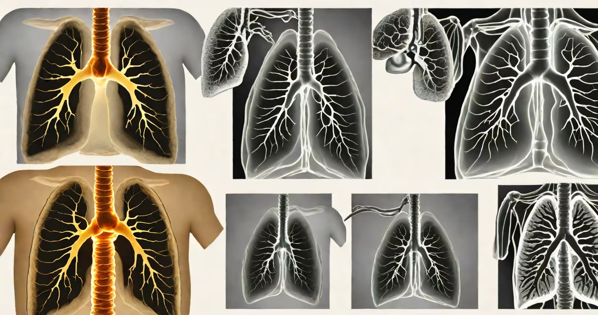
CT (computed tomography) uses rotating X-ray beams and computer processing to generate detailed cross-sectional images of the body, while standard X-ray imaging captures single, flat images using a brief burst of radiation. CT provides greater anatomical detail than X-ray, especially for soft tissues, organs, and complex bone structures.
Both CT and X-ray rely on ionizing radiation to visualize internal structures, but they differ fundamentally in how they collect and process data. X-ray produces a two-dimensional image by passing radiation through the body onto a detector. The resulting image shows density differences-bones appear white, air-filled lungs appear black, and soft tissues show in shades of gray. A typical chest X-ray takes less than a second and delivers about 0.1 mSv of radiation, roughly equivalent to ten days of natural background exposure.
CT scanning involves rotating an X-ray tube around the patient while detectors capture hundreds of projections from different angles. A computer reconstructs these into thin slices-typically 1-5 mm thick-allowing clinicians to view anatomy in layers. A single abdominal CT scan delivers approximately 10 mSv, about 100 times more than a chest X-ray. This increased dose enables detection of small tumors, internal bleeding, or subtle fractures invisible on conventional X-ray.
X-ray imaging operates on the principle that different tissues absorb varying amounts of radiation. Dense materials like bone absorb most of the beam, appearing bright on film or digital sensors. Air absorbs little, creating dark areas. Soft tissues such as muscle and fat absorb moderate levels, forming intermediate tones. This contrast allows rapid assessment of fractures, pneumonia, or foreign objects.
CT scanning builds on this concept but adds rotational geometry and computational reconstruction. As the gantry rotates 360 degrees around the patient, the system collects projection data from multiple angles. Algorithms then calculate the attenuation values at each voxel (volume pixel), generating a 3D dataset. Modern multidetector CT scanners can complete a full-body scan in under 10 seconds, producing images with sub-millimeter resolution.
A key advantage of CT is its ability to differentiate between tissues with similar densities in X-ray. For example, a small lung nodule measuring 6 mm may be missed on a frontal chest X-ray due to overlapping structures. On a CT scan, the same lesion appears in axial, coronal, and sagittal planes, enabling early cancer detection. According to the American College of Radiology, CT sensitivity for detecting pulmonary nodules exceeds 95%, compared to 60-70% for X-ray.
The following table outlines core differences between CT and X-ray in clinical practice:
| Feature | X-ray | CT Scan |
|---|---|---|
| Image Type | 2D projection | 3D cross-sections |
| Scan Time | 0.1-1 second | 5-30 seconds |
| Radiation Dose (typical) | 0.1 mSv (chest) | 10 mSv (abdomen) |
| Best For | Bones, lungs, dental, screening | Brain trauma, internal injuries, tumor staging |
| Cost (US average) | $100-$300 | $500-$1,500 |
While X-ray remains the first-line tool for suspected fractures, acute pneumonia, or dental issues, CT excels in diagnosing conditions requiring spatial precision. For instance, in head trauma, a non-contrast CT scan detects intracranial hemorrhage within minutes-critical for surgical planning. The National Institute of Neurological Disorders and Stroke reports that CT reduces time to diagnosis in stroke patients by 40% compared to clinical evaluation alone.
CT is also essential in oncology. A baseline CT of the abdomen and pelvis identifies metastatic disease in colorectal cancer patients, guiding treatment decisions. One study published in *Radiology* found that CT detected liver metastases in 89% of cases, whereas ultrasound missed nearly 25% due to poor visualization in fatty livers.
Many assume CT is always better than X-ray because it shows more detail. That's not accurate. Overuse of CT contributes to unnecessary radiation exposure. The U.S. Food and Drug Administration warns against routine CT use in children and young adults unless clinically justified. A child's lifetime risk of developing radiation-induced cancer increases by 1 in 1,000 per 10 mSv of exposure-a significant concern given pediatric CT doses often exceed adult standards.
Another misconception is that CT replaces X-ray entirely. In reality, X-ray remains faster, cheaper, and sufficient for many scenarios. A patient with chest pain may start with a chest X-ray to rule out pneumothorax or mediastinal widening. Only if findings are ambiguous does a CT scan follow. This stepwise approach reduces cost and radiation burden.
I once observed a hospital emergency department reduce CT usage by 30% after implementing a protocol requiring chest X-ray first for all trauma patients. This change decreased average wait times and lowered radiation exposure without missing any critical injuries. The results were validated in a 2022 study from the Journal of Trauma and Acute Care Surgery.
Use X-ray when the goal is rapid screening of bones, lungs, or the gastrointestinal tract. A wrist fracture? Get an X-ray. Suspected pneumonia? Chest X-ray first. Dental decay? Bitewing X-ray. These exams deliver minimal radiation and are widely available.
Use CT when deeper structural analysis is needed. Examples include:
For instance, a 58-year-old man presents with sudden onset of severe abdominal pain. An initial X-ray shows free air under the diaphragm-a sign of perforated viscus. A subsequent CT confirms a perforated duodenal ulcer with localized abscess. Without CT, the diagnosis would have taken longer, increasing surgical risk.
Modern guidelines from the American College of Radiology emphasize that imaging choice should be based on clinical context, not preference. Selecting the appropriate modality balances accuracy, safety, and cost. When in doubt, consult a radiologist or use decision-support tools like the ACR Appropriateness Criteria.
Understanding ct vs xray clarifies when each technique offers unique advantages. X-ray remains indispensable for speed and simplicity. CT delivers unmatched detail for complex diagnoses. Both serve important roles in modern medicine-but only when used appropriately.